Drawing Of Skeletal Muscle
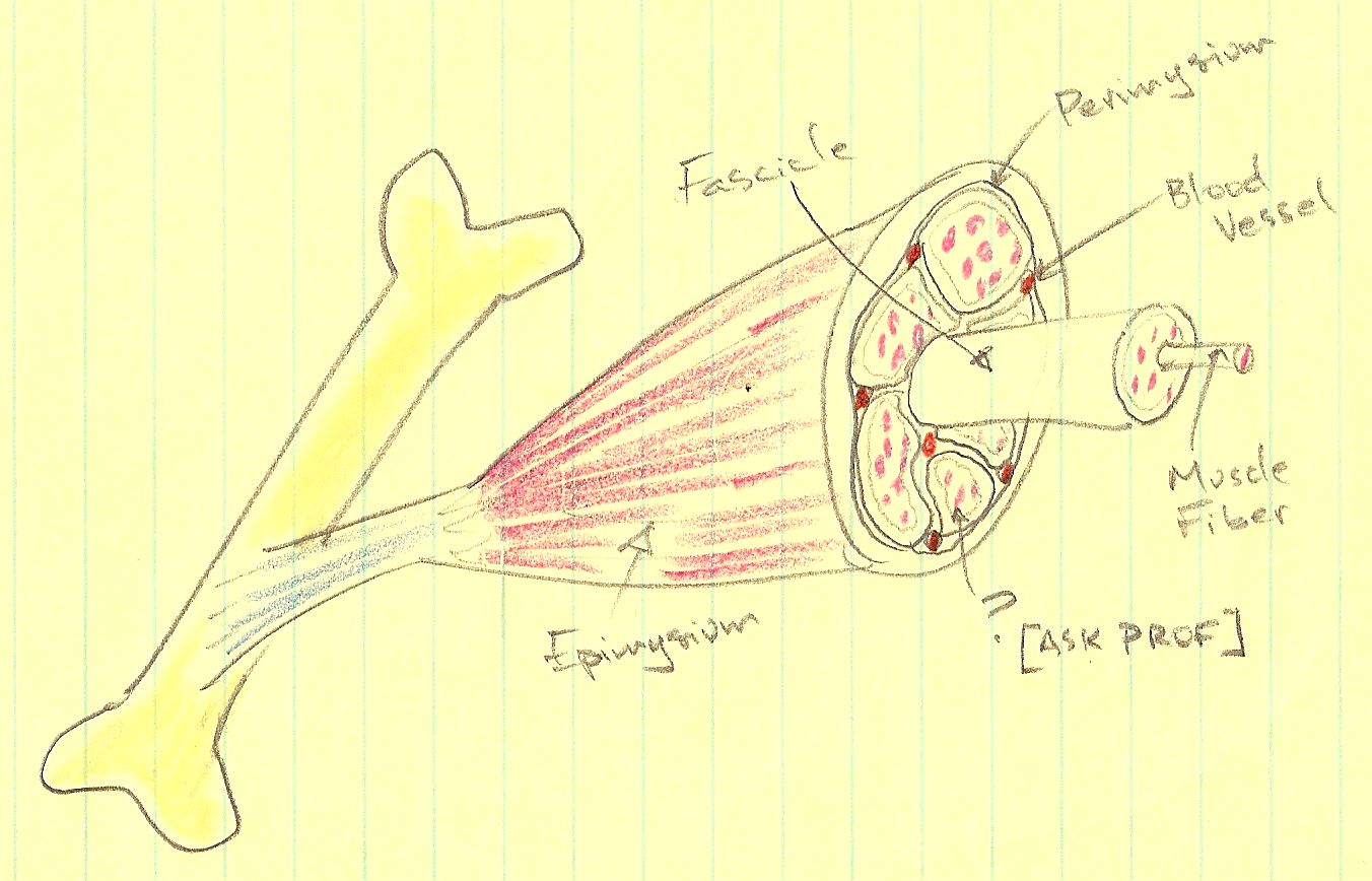
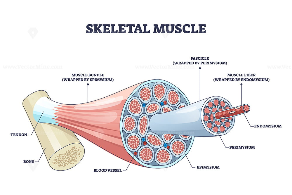
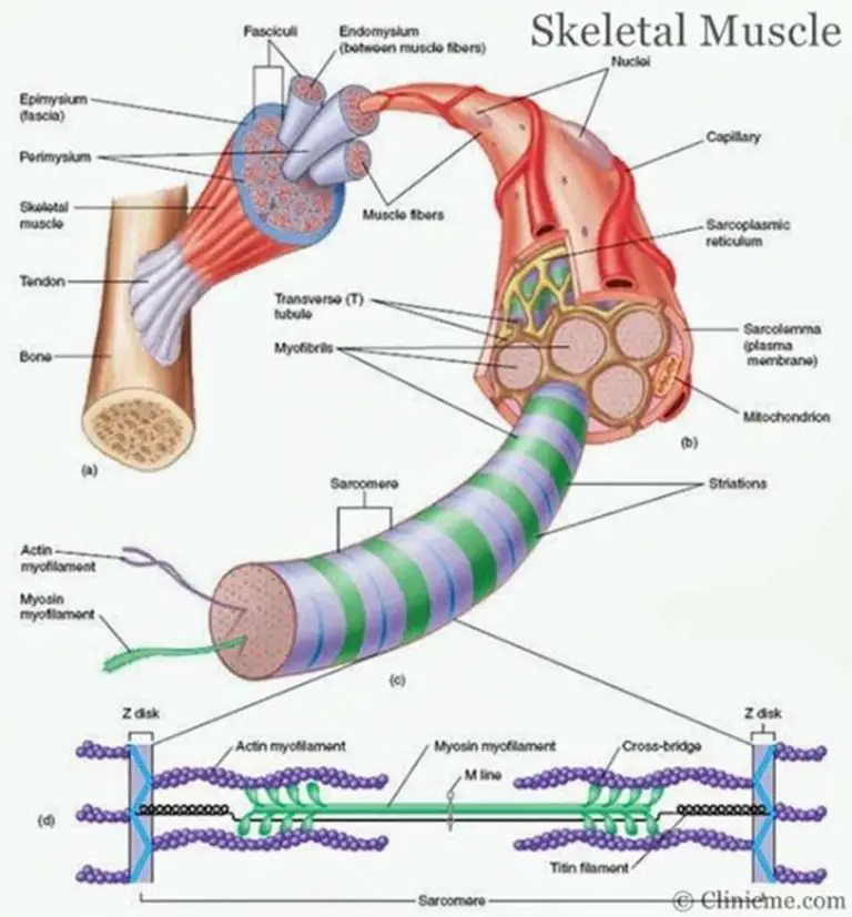
Drawing Of Skeletal Muscle - Web different type of muscle cells have different unique characteristics. The musculoskeletal system (locomotor system) is a human body system that provides our body with movement, stability, shape, and support. It is the pen diagram of skeletal, smooth and cardiac muscle for class 10, 11 and 12. Speaking, walking, or writing) requires skeletal muscle. Each chart groups the muscles of that region into its component groups, making your revision a million times easier. Describe the layers of connective tissues packaging skeletal muscle. It consists of long multinucleate fibers. Explain how muscles work with tendons to move the body. The fibers run the entire length of the muscle they come from and so are usually too long to have their ends visible when viewed under the microscope. A comprehensive guide to drawing realistic muscles. Web these tissues include the skeletal muscle fibers, blood vessels, nerve fibers, and connective tissue. Explain how muscles work with tendons to move the body. This type of muscle creates movement in the body. Within muscles, there are layers of connective tissue called the epimysium, perimysium, and endomysium. Web skeletal muscle is an excitable, contractile tissue responsible for maintaining posture and moving the orbits, together with the appendicular and axial skeletons. Identify areas of the skeletal muscle fibers. Bones are the foundation of the body. A comprehensive guide to drawing realistic muscles. Here, let's learn more about the skeletal muscle description with a labelled diagram. Web identify and describe the microscopic anatomy of a muscle fiber and a sarcomere. Web in the musculoskeletal system, the muscular and skeletal systems work together to support and move the body. By the end of this section, you will be able to: Explain how muscles work with tendons to move the body. Web anatomy of a skeletal muscle cell. The fibers run the entire length of the muscle they come from and so. Identify areas of the skeletal muscle. Speaking, walking, or writing) requires skeletal muscle. A comprehensive guide to drawing realistic muscles. Web skeletal muscle is found attached to bones. They are made of muscle fibres and play an important role in muscle excitation and contraction. Skeletal muscles act not only to produce movement but also to stop movement, such as resisting gravity to maintain posture. Web skeletal muscle is found attached to bones. Each skeletal muscle has three layers of connective tissue that enclose it and provide structure to the muscle as a whole, and also compartmentalize the muscle fibers within the muscle. By the. It consists of long multinucleate fibers. The musculoskeletal system (locomotor system) is a human body system that provides our body with movement, stability, shape, and support. Describe the layers of connective tissues packaging skeletal muscle. These layers cover muscle subunits, individual muscle cells, and myofibrils respectively. A comprehensive guide to drawing realistic muscles. Web practice drawing different muscle groups, including the biceps, triceps, abs, and quads. By the end of this section, you will be able to: Web skeletal muscles contain connective tissue, blood vessels, and nerves. The bones of the skeletal system serve to protect the body's organs, support the weight of the body, and give the body shape. Web skeletal muscles. Understanding the anatomy of muscles is essential for realistic depictions. For example, the skeletal muscle is the only type of muscle cell that is always multinucleated (for more info see the latter half of sal's video). Each skeletal muscle has three layers of connective tissue that enclose it and provide structure to the muscle as a whole, and also compartmentalize. After all, they offer anchor points for muscle origin and insertion. Web a complete list of muscles. Web practice drawing different muscle groups, including the biceps, triceps, abs, and quads. Web skeletal muscles contain connective tissue, blood vessels, and nerves. Web skeletal muscles are voluntary and striated in nature. Web a complete list of muscles. The bones of the skeletal system serve to protect the body's organs, support the weight of the body, and give the body shape. There are three layers of connective tissue: A comprehensive guide to drawing realistic muscles. Web anatomy of a skeletal muscle cell. Web in this video i have shown the simplest way of drawing muscle drawing. It is the pen diagram of skeletal, smooth and cardiac muscle for class 10, 11 and 12. Web skeletal muscles contain connective tissue, blood vessels, and nerves. Web identify and describe the microscopic anatomy of a muscle fiber and a sarcomere. Here are some key points. Skeletal muscle fibers are organized into groups called fascicles. Cardiac muscles, found only in the heart, work involuntarily and at a moderate speed to keep our heart beating. Speaking, walking, or writing) requires skeletal muscle. Mastering the human skeleton will mean you get figure drawing right every time. We’ve created muscle anatomy charts for every muscle containing region of the. Web identify and describe the microscopic anatomy of a muscle fiber and a sarcomere. Web practice drawing different muscle groups, including the biceps, triceps, abs, and quads. Cardiac muscles, found only in the heart, work involuntarily and at a moderate speed to keep our heart beating. Blood vessels and nerves enter the connective tissue and branch in the cell. Web skeletal muscle is an excitable, contractile tissue responsible for maintaining posture and moving the orbits, together with the appendicular and axial skeletons. The musculoskeletal system (locomotor system) is a human body system that provides our body with movement, stability, shape, and support. This type of muscle creates movement in the body. It attaches to bones and the orbits through tendons. Describe the layers of connective tissues packaging skeletal muscle. These layers cover muscle subunits, individual muscle cells, and myofibrils respectively. Web these tissues include the skeletal muscle fibers, blood vessels, nerve fibers, and connective tissue. Each chart groups the muscles of that region into its component groups, making your revision a million times easier. Explain how muscles work with tendons to move the body. Web anatomy of a skeletal muscle cell. Web in the musculoskeletal system, the muscular and skeletal systems work together to support and move the body. Muscles work on a macro level, starting with tendons that attach muscles to bones.
Skeletal Muscle Cell Structure

How To Draw Skeletal, Smooth and Cardiac Muscle Diagram Types Of

Skeletal muscle description with cross section structure outline
Skeletal Muscles Skeletal Muscle Definition DK Find Out

(A) Illustration of skeletal muscle structure copied with permission
Skeletal Muscle Drawing at Explore collection of

Muscle Tissue Drawing at GetDrawings Free download
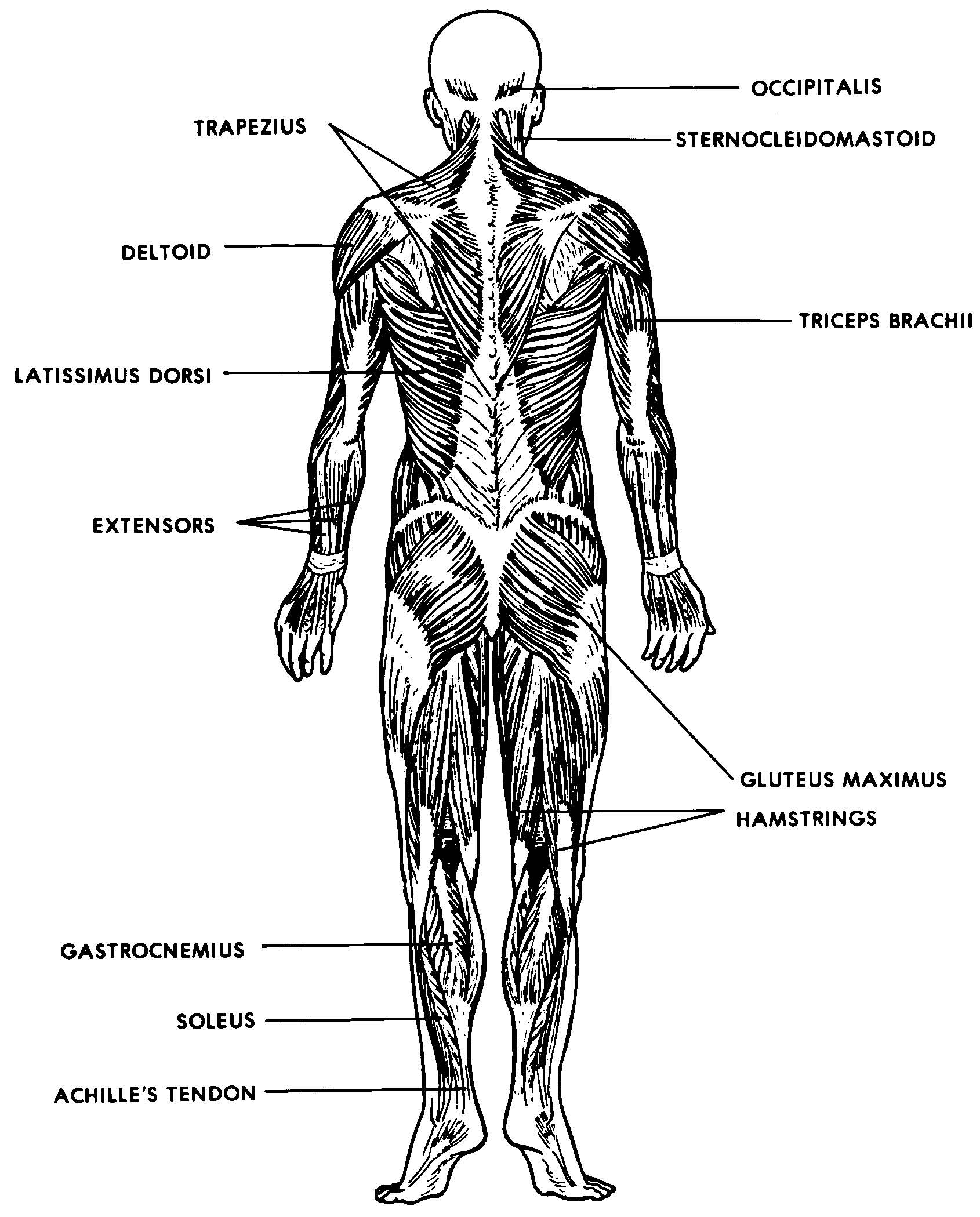
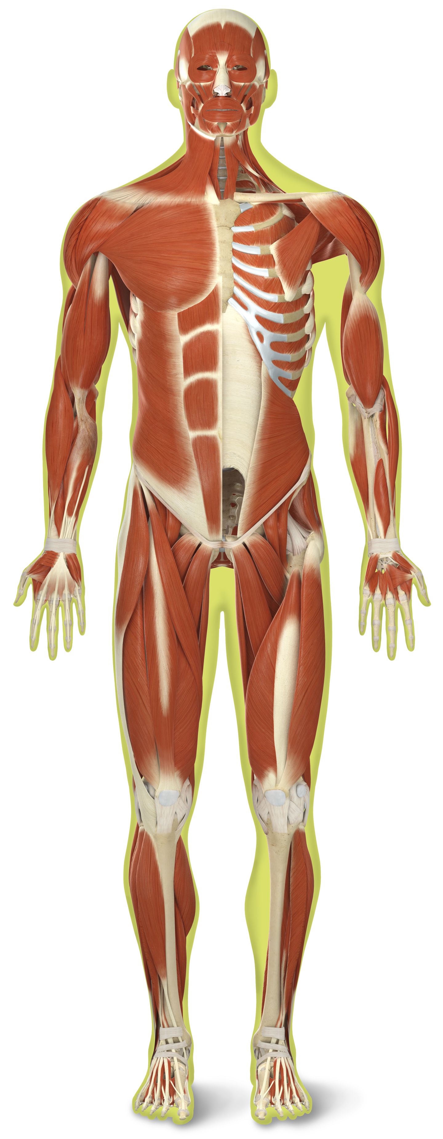
Images 05. Muscular System Basic Human Anatomy
Skeletal muscle structure with anatomical inner layers outline diagram
Skeletal muscle diagram
There Are Three Layers Of Connective Tissue:
Web Different Type Of Muscle Cells Have Different Unique Characteristics.
Skeletal Muscles, Attached To Bones And Tendons, Help Us Move Voluntarily And Quickly.
Web Skeletal Muscle Is Found Attached To Bones.
Related Post: